Function and form are deeply intertwined in biology. Understanding the physical structures of organisms is crucial to knowing how they grow, adapt, and reproduce. This is where the microscope has played a transformative role in scientific discovery over the past four centuries.
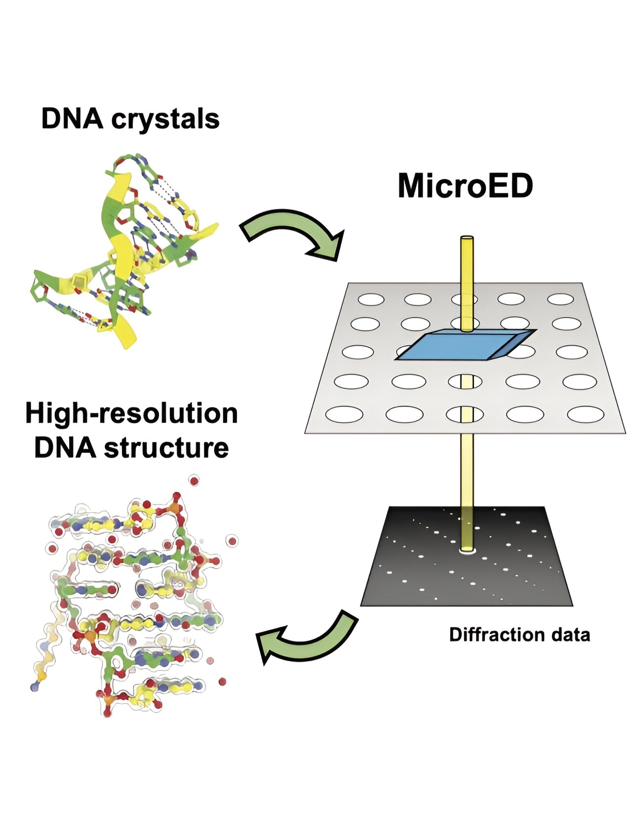
Microscopy, the field of microscope use, has advanced to the point where it can reveal the tiniest of structures. Techniques like microcrystal electron diffraction, or MicroED, allow us to bombard crystalline samples with electrons to obtain detailed information about their atomic configuration.
“The method was initially developed to solve the structure of proteins,” explained Brent Nannenga, an associate professor of chemical engineering at Arizona State University. “But researchers started applying it to pharmaceutical compounds and small organic molecules. The question was whether it would work for DNA.”
Nannenga and his colleagues have now answered that question. They successfully demonstrated the first use of MicroED to analyze a DNA crystal, a breakthrough published in the journal Structure.
For decades, scientists have used X-ray crystallography to study crystal structures. However, this method requires relatively large crystals, which can be challenging to produce in a laboratory and can slow down research progress.
“We’re talking about crystals that are dozens or hundreds of microns in size,” said Nannenga. “But with MicroED, we can work with material that is just hundreds of nanometers, which is a fraction of a single micron.”
MicroED allows researchers to work with smaller, easier-to-produce crystals. However, even these tiny crystals may still be too big for the technique. To overcome this, a process called cryo-focused ion beam (cryo-FIB) milling is used to reduce the crystals to an ideal size of 200 to 250 nanometers.
By combining cryo-FIB milling and MicroED, high-resolution data can be obtained for crystal structure determination. While previous studies have shown the effectiveness of this technique for analyzing biomolecular systems, Nannenga and his team are the first to demonstrate its potential for determining nucleic acid structure.
This approach opens up new opportunities to understand not only DNA but also the structure and function of RNA. These discoveries can support the development of novel nanotechnologies and more effective pharmacological treatments.
Other authors of the paper include Alison Haymaker and Andrey Bardin from Arizona State University, as well as Tamir Gonen and Michael Martynowycz from the University of California, Los Angeles.