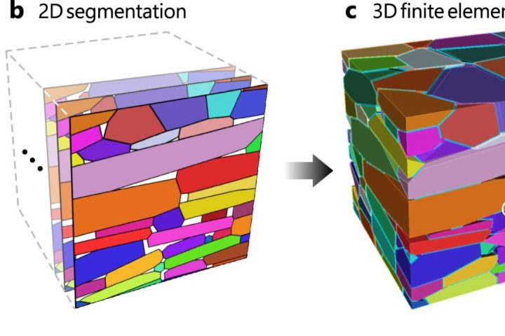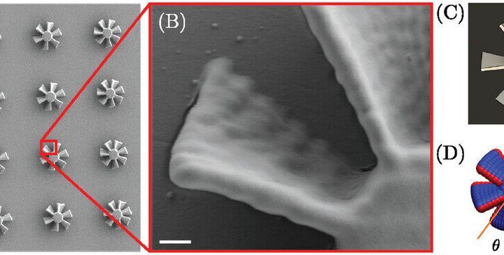
A groundbreaking discovery has been made by a team of bioengineers and biomedical scientists from the University of Sydney and the Children’s Medical Research Institute (CMRI) at Westmead. They have successfully used 3D photolithographic printing to create a complex environment for assembling tissue that mimics the architecture of an organ.
The team, led by Professor Hala Zreiqat and Dr. Peter Newman at the University of Sydney’s School of Biomedical Engineering, along with developmental biologist Professor Patrick Tam from CMRI’s Embryology Research Unit, published their findings in Advanced Science.
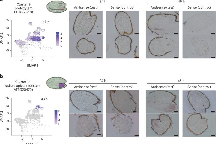
Using bioengineering and cell culture methods, they were able to instruct stem cells to become specialized cells that can assemble into an organ-like structure. This process involves strategically positioned proteins and mechanical triggers that mimic developmental processes.
Professor Hala Zreiqat explained, “Our new method serves as an instruction manual for cells, allowing them to create tissues that are better organized and more closely resemble their natural counterparts. This is an important step towards being able to 3D print working tissue and organs.”
Dr. Peter Newman compared building tissues from cells to constructing a building from many different parts. He said, “Imagine trying to build a Lego castle by randomly scattering the blocks on a table and hoping that they’ll fall into the correct place. Even though each block is designed to connect with others, without a clear plan, you’d likely end up with something that looks more like a large pile of disconnected Lego blocks rather than a castle.”
Seven News coverage
Dr. Newman further explained, “The same can be said about building organs and tissues from cells: without specific instructions, the cells would likely group together unpredictably within the incorrect structures. What we’ve effectively done is create a step-by-step process that guides each building block to exactly where it should go and how it should connect with the others.”
“In line with this approach, our recently published work applies a new 3D printing method to define instructions for cells that guide them into forming more organized and accurate structures. Through this, we’ve created a bone-fat assembly that resembles the structure of bone and an assembly of tissues that resemble processes during early mammalian development.”
This research into complex tissue and organ-like structures, known as organoids, not only helps us understand how organs develop and function, but also provides insights into how genetic mutations and developmental errors can cause diseases. It also opens up possibilities for cell and gene therapy.
Professor Hala Zreiqat emphasized the practical implications of this method, saying, “Beyond understanding the intricate ‘instruction manual’ of life, this method has immense practical implications. For instance, in regenerative medicine, where there is a pressing need for organ transplants, further research using this approach may facilitate the growth of functional tissues in the lab. Imagine a future where the waitlist for organ transplants could be drastically reduced because we can generate such tissues in the lab that sufficiently resemble their natural counterparts.”
Dr. Newman added, “Moreover, this technology could revolutionize how we study and understand diseases. By creating accurate models of diseased tissues, we can observe disease progression and treatment responses in a controlled environment. We hope this could one day lead to more effective treatments and even cures for diseases that are currently hard to tackle.”
Professor Patrick Tam from CMRI highlighted the significance of this bioengineering technology, stating, “In the past, stem cells were grown to generate many cell types, but we could not control how they differentiate and assemble in 3D. With this bioengineering technology, we can now direct the stem cells to form specific cell types and organize these cells properly in time and space, thereby recapitulating the real-life development of the organ.”
The researchers are optimistic about the potential of their research in treating vision loss caused by conditions such as macular degeneration and inherited diseases affecting retinal photoreceptor cells.
Professor Tam explained, “If we can generate a patch of cells by bioengineering and see how the whole system functions, then we can investigate therapies that use functional cells to replace cells in the eye that were lost because of disease. It would have great impact if we can deliver healthy cells into the eye. Regardless of whether the macula had been lost due to inherited disease or trauma, the treatment would be the same.”
“The idea of treating rare genetic diseases and improving quality of life in this way is empowering. We expect that this work will lead to advanced therapies that can be moved into practice.”
The team’s next focus is to further develop this technique and explore its potential in regenerative medicine and new treatment approaches for various diseases.