A new way of looking at the brain
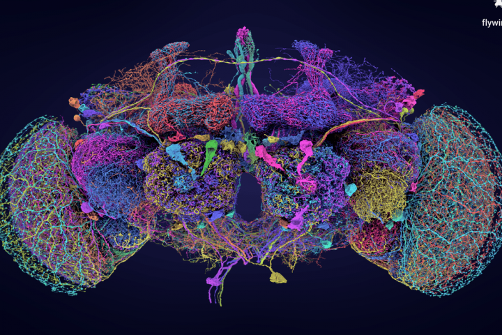
Prepare to have your mind blown. Functional magnetic resonance imaging, or fMRI, has revolutionized brain research. Developed in the 1990s, this groundbreaking technique allows scientists to measure blood oxygenation in different parts of the brain while a person is performing a task or being exposed to a stimulus.
Here’s how it works: brain areas with higher activity demand more oxygen, resulting in increased blood flow to those areas. So, imagine lying inside a big tube for an hour, pushing buttons and listening to a researcher’s voice while trying not to move your head. The result? A mind-blowing 3D map of your brain, color-coded to show where your neurons are firing the most.
Because fMRI is noninvasive and safe, it has become a staple in neuroscience research. It has transformed our understanding of how the brain works, shattered misconceptions, and holds the potential to read our thoughts with the help of AI technology in the future.
But, there’s a catch. And brace yourself, because it involves math.
The issue of multiple comparisons
Scientists aren’t just scanning brains for fun; they’re trying to answer important questions. To do that, they need to perform statistical analysis. However, when you have a lot of data and run numerous statistical tests, there’s a risk of finding false positives by chance alone. So, how do you avoid this pitfall?
Enter multiple comparisons correction, a collection of methods developed by statisticians to address this very problem. fMRI studies generate vast amounts of data, which undergo multiple statistical tests to compare different variables. Within this process, false positives can occur, but correcting too harshly can lead to false negatives. It’s a delicate balance.
This issue caught the attention of lead study author Craig Bennett, and it all started with a salmon.
“We should scan a whole fish”
Now, let’s dive into the salmon story. Bennett recounts how, as a grad student, he and his colleagues wanted to test their new MRI protocols with interesting objects. They began with a pumpkin, then moved on to a Cornish game hen. But they decided to up the ante and exclaimed, “We should scan a whole fish!”
Early one Saturday morning in 2005, Bennett purchased a full-length Atlantic salmon from a local supermarket. The cashier was understandably surprised by the purchase and even more so when Bennett explained what was about to happen to the fish. The salmon was then scanned while being shown images of human faces and, unsurprisingly, performed poorly due to being dead. After the scan, the salmon was reportedly consumed, and the data was filed away.
Fast forward to 2008, when a discussion about multiple comparisons correction led Bennett to revisit the salmon scans. What he discovered left him astounded. Despite being dead, the salmon’s brain showed a cluster of significant voxels along its midline, suggesting that it was “thinking” about the images it was shown during the scan.
Initially met with skepticism, Bennett and his colleagues eventually presented their findings at a conference, and the significance of the salmon’s brain activity became clear. A full paper was published, and the rest is history.
Where are we now?
Since the infamous dead salmon study, the debate surrounding how to report and interpret fMRI data has only intensified. It’s important to note that the study was never meant to discredit fMRI itself; the technique remains a valuable tool in neuroscience. However, it has sparked a necessary conversation about how to handle the data it produces.
fMRI studies continue to yield fascinating discoveries, many of which involve living animals. Despite the hilarity surrounding the dead salmon, it has played a significant role in the field’s development, even earning the team an Ig Nobel Prize in 2012.
As Bennett himself concluded, “The fish has the chance to impact the field of neuroimaging in a very positive way.” We can only imagine how proud the salmon would be of its unexpected legacy.