Zeolites have unique porous atomic structures and are incredibly versatile, serving as catalysts, ion exchangers, and molecular sieves. However, due to their low electron irradiation resistance, it has been challenging to directly observe the local atomic structures of zeolites using electron microscopy. As a result, the fundamental property-structure relationships of these materials have remained unclear.
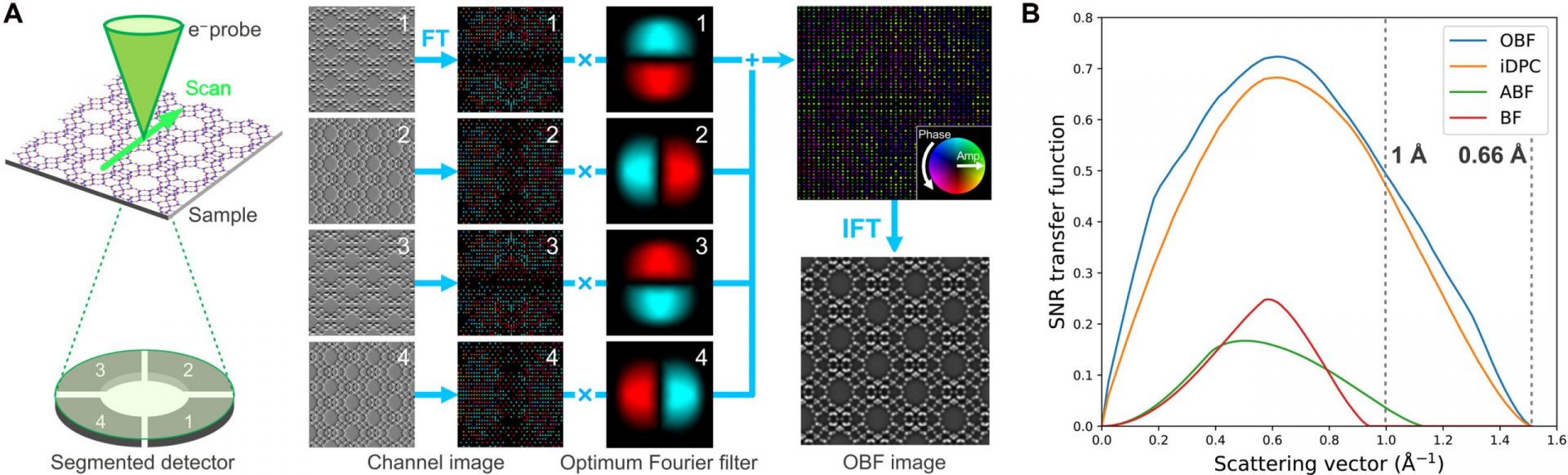
Recent advancements in imaging technology have introduced a low-electron dose imaging method called optimum bright-field scanning transmission electron microscopy (OBF STEM). This method allows for the reconstruction of high signal-to-noise ratio images with high dose efficiency.
In a groundbreaking study, Kousuke Ooe and a team of scientists from the University of Tokyo and the Japan Fine Ceramics Center utilized OBF STEM to observe the atomic structures of two types of zeolites. By examining the complex atomic structure of a faujasite-type (FAU) zeolite, they were able to gain insights into the local atomic structures of electron beam-sensitive materials.
Analyzing zeolites in the materials lab
Zeolites are remarkable porous materials that possess regularly arranged nanosized pores, making them ideal for various applications in catalysis, gas separation, and ion exchange. The properties of zeolites are closely tied to their pore geometry, which allows for interactions with guest molecules and ions. Traditionally, diffractometric methods have been used to analyze the structure of zeolites.
While scanning electron microscopy has proven to be a powerful tool for analyzing local structures and observing the atomic arrangement of electron-resistant materials at the sub-angstrom level, zeolites are more electron-beam sensitive compared to other organic materials. This sensitivity limits the use of electron microscopy-based observations due to electron irradiation.
Optimum bright-field scanning transmission electron microscopy (OBF/STEM)
In 1958, materials scientist J. W. Menter used a high-resolution transmission electron microscope to observe zeolites and reported a lattice resolution of 14 Angstrom. Advanced imaging techniques in the 1990s significantly improved the visualization of zeolite frameworks, but observing atomic sites within the materials remained challenging.
Recent advancements in scanning transmission electron microscopy (STEM) electron detectors have led to the development of more advanced imaging methods, such as the optimum bright-field (OBF) STEM method. This method allows for the observation of atomic structures with the highest signal-to-noise ratio in real-time.
In their study, Ooe and his colleagues utilized real-time OBF imaging to determine the architecture of zeolites at subangstrom resolution. The results highlighted the capability of advanced electron microscopy to characterize the local structure of beam-sensitive materials.
Direct imaging of atomic structures in zeolites: Real-time OBF imaging vs. STEM imaging
The zeolite framework consists of two building blocks: sodalite cages and double 6-membered rings. Using real-time optimum bright-field (OBF) imaging, the team successfully detected the framework of the material. By using an electron probe current of 0.5 pico-angstrom to prevent beam-related damage, they were able to analyze the typical inorganic materials. They then compared the OBF images with other scanning transmission electron microscopy images obtained under similar dose conditions.
While the existing STEM methods provided a basic structure of the material framework, analyzing the atomic structure using this method proved challenging due to low current dosage. In contrast, the OBF images offered more reliable and interpretable image contrast with higher dose efficiency.
Direct observation of the twin boundary
The research team used the optimum bright-field method to examine the atomic structure of a twin boundary in the zeolite structure. The framework was created by stacking a layered structure unit known as a “faujasite sheet” in a cubic manner. The OBF imaging results showed a power spectrum of the image with an information transfer beyond 1 Angstrom. The low-dose light-element imaging with OBF STEM provided a better alternative for analyzing the structure of zeolites, including the local change of symmetry.
Ooe and his colleagues conducted density functional theory calculations to examine the stability of the twin boundary structure, and the experimental image agreed with the simulated counterpart. They also applied the method to a different type of zeolite sample, demonstrating the importance of the silicon aluminum ratio in influencing the material properties and the adherence of ions and molecules. The atomic observations of a sodium-based zeolite sample using this method facilitated the identification of extra cation sites with low occupancy in the zeolitic framework.
Outlook
In summary, Kousuke Ooe and his colleagues have developed a dose-efficient scanning transmission electron microscopy imaging method called “optimum bright field scanning transmission electron microscopy” (OBF-STEM) for low-dose atomic resolution imaging. This method allows for the direct visualization of atomic structures in faujasite-type zeolite materials, which are known for their sensitivity to electron beams. The method can also be applied to other beam-sensitive materials to characterize their local atomic structures and study structure-property relationships.