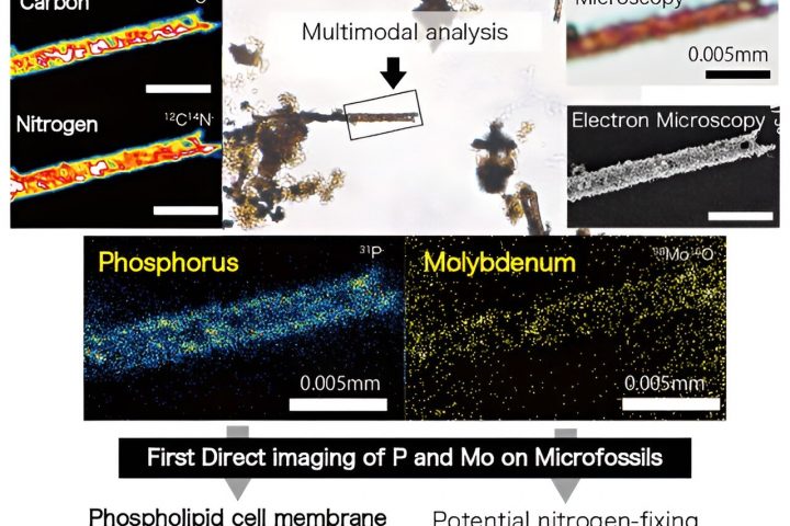
Exciting breakthrough in imaging technology! Researchers have developed a cutting-edge algorithm that revolutionizes the way we recover the 3D refractive index distribution of biological samples. This algorithm is a game-changer for a new imaging approach called intensity diffraction tomography (IDT).
Get ready for the future of biomedical imaging! Jiabei Zhu from Boston University will be presenting this groundbreaking research at the Optica Imaging Congress, happening from 14—17 August 2023 in Boston, Massachusetts.
“3D quantitative phase imaging (QPI) has opened up endless possibilities in the field of biomedical imaging. With QPI, we can now capture detailed images of transparent living organisms and cells without the need for contrast agents or dyes that can harm the sample,” explains Zhu.
“QPI goes beyond traditional microscopy techniques by not only providing high-contrast morphological information but also quantitative phase information. In particular, 3D QPI allows us to visualize the high-resolution 3D refractive index (RI) distribution inside the samples. This invaluable information greatly aids research in hematology, neurology, and immunology, and enhances disease and infection diagnosis.”
However, achieving high-speed acquisition and high resolution in 3D imaging of thick biological samples has always been a challenge. That’s where IDT comes in. IDT is a label-free phase tomography technique that overcomes these limitations. It can be easily integrated into a standard microscope using a programmable LED array.
Zhu’s research team has recently developed two groundbreaking IDT methods: annular IDT (aIDT) and multiplexed IDT (mIDT). These methods have significantly boosted image acquisition speed, allowing for the visualization of dynamic biological samples. aIDT uses an LED ring that matches the objective’s numerical aperture, while mIDT uses multiple LEDs to simultaneously illuminate the sample.
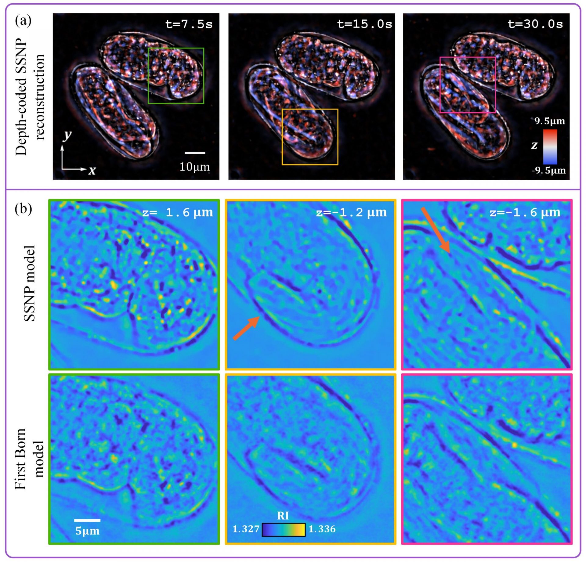
However, the researchers encountered a challenge when existing IDT reconstruction algorithms failed to work effectively with their new approaches, which involved high-NA objectives. Undeterred, they decided to develop a brand new algorithm. This algorithm utilizes a multiple scattering model based on the split-step non-paraxial (SSNP) method, which was recently developed to overcome similar limitations in optical diffraction tomography.
The researchers conducted experiments using buccal epithelial cells with aIDT and were amazed by the results. The new IDT reconstruction algorithm allowed for easy discrimination of cells at different depths, reconstruction of cell boundaries and membranes, and visualization of native bacteria surrounding the cells.
They also applied the algorithm to a thick multi-scattering live C. elegans embryo using mIDT. The resulting reconstructed images revealed intricate details of how the worms were folded, and a single-depth cross-section provided morphological insights into the cells’ outline, the buccal cavity, and the tail of the worm.
Overall, these experiments demonstrated that by extending the SSNP method to IDT, the researchers achieved high-quality images with a large field of view. This breakthrough has the potential to revolutionize the field of biomedical imaging and open up new avenues for research and diagnosis.